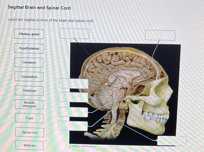
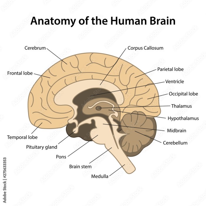
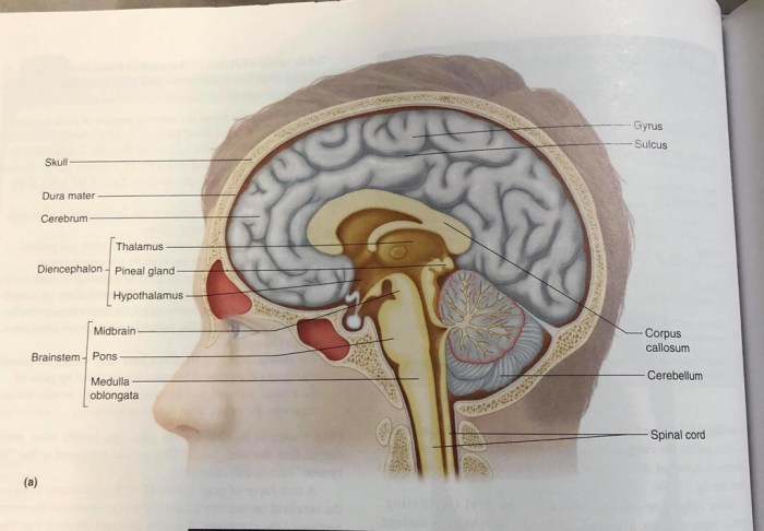
Label the sagittal section of the brain and spinal cord – Labeling the sagittal section of the brain and spinal cord is a crucial step in understanding the intricate anatomy of the central nervous system. This comprehensive guide provides a detailed overview of the structures visible in these sections, the imaging techniques used to visualize them, and their clinical and research applications.
The sagittal section, a midline view of the brain and spinal cord, reveals the complex arrangement of neural structures that govern our thoughts, actions, and bodily functions. Understanding the anatomy of this section is essential for neurosurgeons, neurologists, and researchers seeking to diagnose and treat neurological disorders.
1. Introduction

The sagittal section of the brain and spinal cord provides a medial view of the central nervous system, revealing its intricate structures and their relationships. Understanding this section is crucial for comprehending the anatomy, function, and pathology of the brain and spinal cord.
2. Anatomy of the Sagittal Section

Brain
The sagittal section of the brain displays the cerebrum, cerebellum, brainstem, and ventricles. The cerebrum, the largest part of the brain, is responsible for higher-order cognitive functions. The cerebellum, located behind the cerebrum, coordinates movement and balance. The brainstem, connecting the cerebrum and cerebellum, controls vital functions such as breathing and heart rate.
The ventricles are fluid-filled cavities within the brain that produce and circulate cerebrospinal fluid.
Spinal Cord
The sagittal section of the spinal cord reveals its segmentation into 31 segments, each giving rise to a pair of spinal nerves. The spinal cord is composed of gray matter, which contains cell bodies, and white matter, which contains myelinated axons.
3. Imaging Techniques

MRI
Magnetic resonance imaging (MRI) uses strong magnetic fields and radio waves to produce detailed images of the brain and spinal cord. MRI is non-invasive and provides excellent contrast between different tissues, making it valuable for diagnosing and monitoring various neurological conditions.
CT, Label the sagittal section of the brain and spinal cord
Computed tomography (CT) uses X-rays and computer processing to create cross-sectional images of the brain and spinal cord. CT is faster than MRI but provides less detailed images. It is often used for trauma evaluation and detecting bone abnormalities.
Ultrasound
Ultrasound uses high-frequency sound waves to produce images of the brain and spinal cord. Ultrasound is portable and relatively inexpensive, making it useful for point-of-care evaluations.
4. Clinical Applications
The sagittal section of the brain and spinal cord is essential in clinical practice for:
- Diagnosing neurological conditions such as stroke, tumors, and infections.
- Assessing the severity of spinal cord injuries.
- Guiding surgical interventions for brain and spinal cord disorders.
- Monitoring the progression of neurological diseases.
Helpful Answers: Label The Sagittal Section Of The Brain And Spinal Cord
What are the key structures visible in the sagittal section of the brain?
The sagittal section of the brain reveals the cerebrum, cerebellum, brainstem, and ventricles, providing a comprehensive view of the brain’s major components.
How is the sagittal section of the spinal cord used in clinical practice?
The sagittal section of the spinal cord is invaluable for diagnosing and treating spinal cord injuries, tumors, and other neurological conditions, enabling clinicians to visualize the extent of damage and plan appropriate interventions.